
The fetus’s heart begins to beat as early as the fifth week of pregnancy, well before ultrasound allows us to observe its movements. Anomalies can occur at every stage of this development, sometimes without detectable symptoms before birth. In utero cardiac arrests remain rare but require increased monitoring during pregnancy.
Early identification of heart defects significantly improves management while reducing risks for the unborn child. Regular follow-up and targeted examinations are essential to detect any anomalies and anticipate potential complications.
Related reading : Everything You Need to Know About Drink Prices on Cruise Ships in Egypt
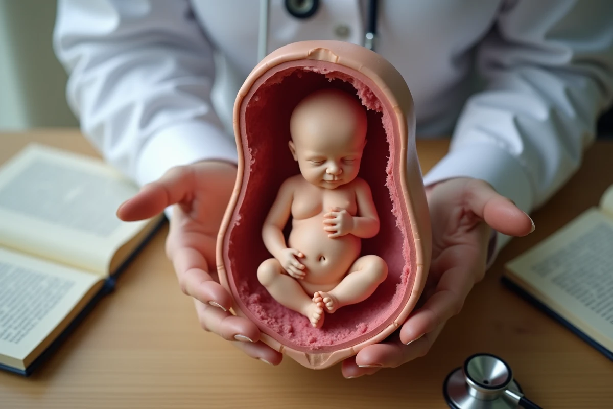
The Fetus’s Heart: Understanding Its Early Stages of Development
Under the microscope of biology, the mechanics of the heart start working without waiting for any external signal. Three weeks after fertilization, two endocardial tubes merge to outline the first contours of what will be the vital engine of the future child. Not a sound, not a sensation yet, but already, blood circulates in an embryo that is organizing at a dizzying speed.
Where it all begins, every detail matters. The ventricles take shape, separated by thin partitions. The valves make their entrance, true control gates of blood flow. Little by little, this blueprint thickens: aorta, pulmonary artery, venae cavae, everything interlocks. The coronary system, responsible for supplying the heart muscle, sets up behind the scenes to ensure the robustness of this tiny but vital pump.
Read also : Everything You Need to Know About the Other Carrier and Package Tracking for Your Deliveries
By the seventh week, it is no longer just a simple tube, but an almost fully developed heart. The fetus’s heart in utero now orchestrates the supply of oxygen and nutrients. Behind this evolution lies a precise sequence of growth, remodeling, synchronization: each contraction prepares for future autonomy. For those wishing to explore this topic in more detail or learn about in utero cardiac arrests, the resource “Fetus’s Heart: Understanding In Utero Cardiac Arrests – Parenthèses Bien-être” offers additional insights.
Here are the main stages that mark the formation of the heart in the fetus:
- Cardiac organogenesis: fusion of endocardial tubes, organization of the atria and ventricles
- First beats: detectable activity from the fifth week
- Development of valves and vessels: gradual establishment of intrauterine blood circuits
What are the main cardiac anomalies detected before birth?
Fetal echocardiography has completely transformed the ability to spot heart anomalies from the earliest stages of pregnancy. Today, it is not uncommon to identify, sometimes before the end of the first trimester, malformations that will disrupt family journeys and guide medical management. The numbers are stubborn: nearly 1% of births are affected by cardiac anomalies, ranging from simple defects to complex surgical situations.
Among the most frequently observed cases, abnormal communications between the heart chambers, whether inter-ventricular or inter-atrial, alter the natural flow of blood and impose an additional effort on the heart. Other situations, such as transposition of the great vessels, pulmonary atresia, or certain anomalies of the ductus arteriosus, draw attention as soon as diagnosed. These conditions often require specific organization from birth, sometimes even rapid intervention.
Monitoring does not stop at structure: the fetal heart rate is closely scrutinized. Bradycardia, tachycardia, or abnormal variations are monitored through fetal monitoring. An atypical rhythm may signal an acute problem or an underlying genetic condition, such as certain trisomies often associated with cardiac malformations. Antenatal detection allows for adapting birth preparation, anticipating interventions, and reducing the risks of serious complications.
To better understand the types of anomalies encountered before birth, we distinguish:
- Structural heart defects: partition defects, transposition, atresia
- Heart rhythm anomalies: bradycardia, tachycardia, absence of variability
- Signs associated with genetic or chromosomal syndromes: trisomies, genetic anomalies
Prenatal Follow-Up: How Future Parents Can Support Their Baby’s Heart Health
The baby’s heart health is built well before the first cry. At each appointment, the midwife or obstetrician-gynecologist checks the frequency and variability of the heart rate, scrutinizing the slightest atypical signs. This follow-up allows for the rapid detection of rhythm disorders or the absence of variability, sometimes discreet signals of distress or anomalies to explore.
At each stage, different examinations complement the monitoring: ultrasounds, labor monitoring, control of the umbilical cord. Parents are involved in this process: they ask questions, learn about heart development, and discuss each result. The regular listening to the fetal heart rate, the variations observed during movements and rest phases become valuable indicators, closely monitored by the entire medical team.
This constant dialogue between healthcare professionals and families makes all the difference. Trust builds, understanding grows. Monitoring fetal heart health means moving forward together, appointment after appointment, towards a birth marked by vigilance and preparation. With each beat, a silent promise: that of a future to build, with the heart firmly anchored.